Sketch And Label Of A Cross Section Of A Long Bone / Chapter 31 Drawings For The Frcs Tr Orth Musculoskeletal Key
Sketch And Label Of A Cross Section Of A Long Bone / Chapter 31 Drawings For The Frcs Tr Orth Musculoskeletal Key. The large dark spots are passages for blood vessels and nerves. Draw a cross section of compact/osteon bone labeling all microscopic structures. Complete figure 6.1a by labeling compact bone and spongy bone. This photo shows a cross section through bone. The anterior bone of the forehead (frontal bone) contains a large cavity (frontal sinus).
(d) undecalcified section subjected to the von kossa method for calcium detection . Draw a cross section of compact/osteon bone labeling all microscopic structures. A long bone has two parts: Polyps can live individually (like many mushroom corals do) or in large colonies that comprise an entire reef structure. The anterior bone of the forehead (frontal bone) contains a large cavity (frontal sinus).
Sketch And Label Of A Cross Section Of A Long Bone Cross Section Of Right Kidney Photograph By Science Source from i0.wp.com Draw a cross section of compact/osteon bone labeling all microscopic structures. The large dark spots are passages for blood vessels and nerves. The little black spots are osteocytes. The anterior bone of the forehead (frontal bone) contains a large cavity (frontal sinus). This photo shows a cross section through bone. A long bone has two parts: Complete figure 6.1a by labeling compact bone and spongy bone. Osteoblasts are cuboidal cells that are located along the bone surface.
The little black spots are osteocytes.
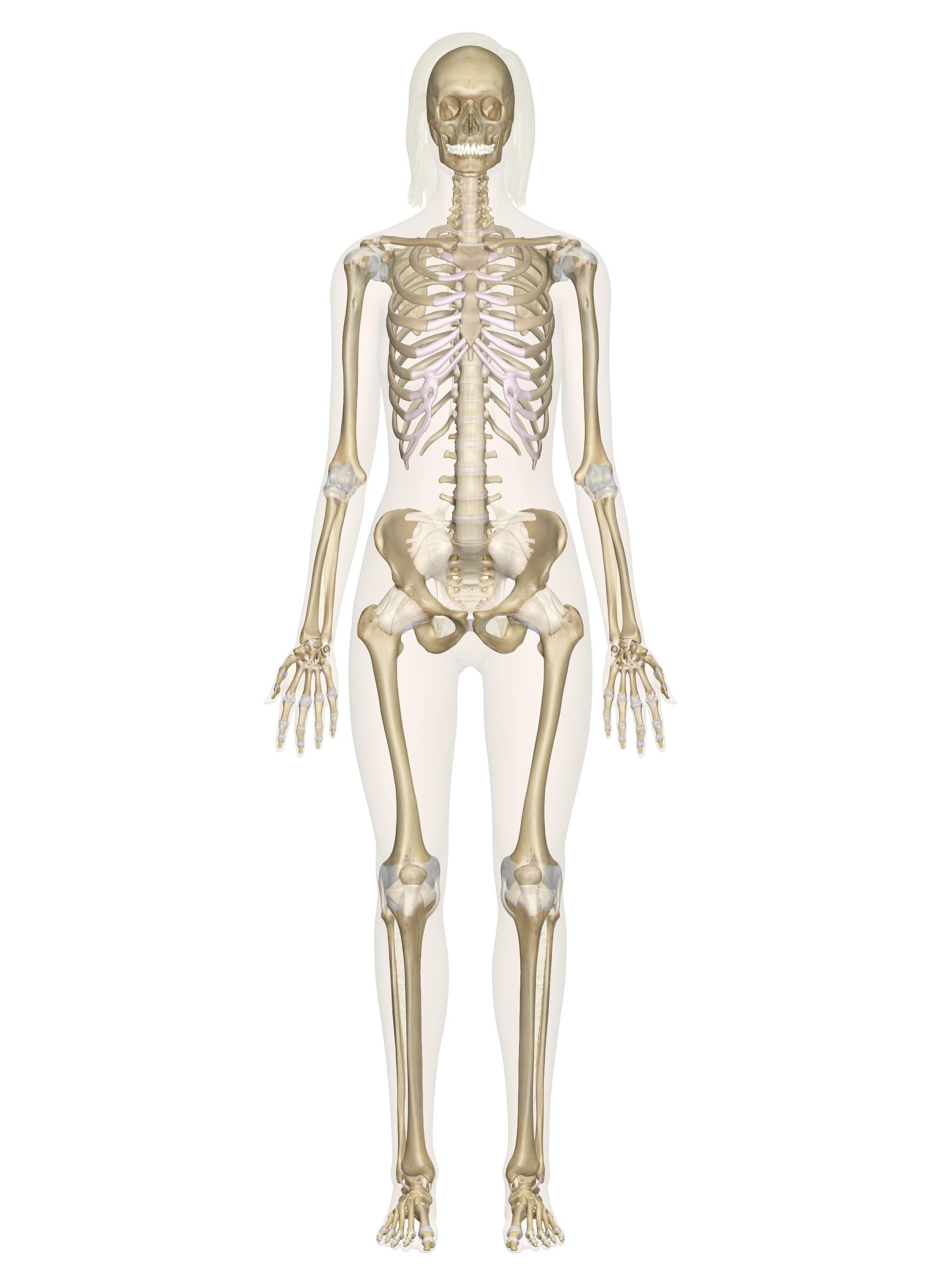
The large dark spots are passages for blood vessels and nerves. The anterior bone of the forehead (frontal bone) contains a large cavity (frontal sinus). Complete figure 6.1a by labeling compact bone and spongy bone. The little black spots are osteocytes. Some, mostly older, compact bone is remodelled to form these haversian systems (or osteons). Draw a cross section of compact/osteon bone labeling all microscopic structures. Polyps can live individually (like many mushroom corals do) or in large colonies that comprise an entire reef structure. A long bone has two parts: Osteoblasts are cuboidal cells that are located along the bone surface. See labeled cross sections of the human body now at kenhub. Long bones, short bones, flat bones, irregular bones, sesamoid. In the long bones, the epiphysis is the region between the growth plate or. The diaphysis and the epiphysis.
Some, mostly older, compact bone is remodelled to form these haversian systems (or osteons). The diaphysis and the epiphysis. (d) undecalcified section subjected to the von kossa method for calcium detection . Long bones, short bones, flat bones, irregular bones, sesamoid. The anterior bone of the forehead (frontal bone) contains a large cavity (frontal sinus).
1 Schematic Drawing Of A Longitudinal Section Through A Long Bone Download Scientific Diagram from www.researchgate.net A long bone has two parts: See labeled cross sections of the human body now at kenhub. Some, mostly older, compact bone is remodelled to form these haversian systems (or osteons). Long bones, short bones, flat bones, irregular bones, sesamoid. This photo shows a cross section through bone. Polyps can live individually (like many mushroom corals do) or in large colonies that comprise an entire reef structure. The large dark spots are passages for blood vessels and nerves. Complete figure 6.1a by labeling compact bone and spongy bone.
See labeled cross sections of the human body now at kenhub.
The large dark spots are passages for blood vessels and nerves. The diaphysis and the epiphysis. Long bones, short bones, flat bones, irregular bones, sesamoid. The little black spots are osteocytes. This photo shows a cross section through bone. In the long bones, the epiphysis is the region between the growth plate or. Some, mostly older, compact bone is remodelled to form these haversian systems (or osteons). Complete figure 6.1a by labeling compact bone and spongy bone. (d) undecalcified section subjected to the von kossa method for calcium detection . See labeled cross sections of the human body now at kenhub. Osteoblasts are cuboidal cells that are located along the bone surface. The anterior bone of the forehead (frontal bone) contains a large cavity (frontal sinus). A long bone has two parts:
Long bones, short bones, flat bones, irregular bones, sesamoid. Draw a cross section of compact/osteon bone labeling all microscopic structures. (d) undecalcified section subjected to the von kossa method for calcium detection . The diaphysis and the epiphysis. This photo shows a cross section through bone.
Skeletal System Labeled Diagrams Of The Human Skeleton from innerbody.imgix.net Complete figure 6.1a by labeling compact bone and spongy bone. Draw a cross section of compact/osteon bone labeling all microscopic structures. Osteoblasts are cuboidal cells that are located along the bone surface. Long bones, short bones, flat bones, irregular bones, sesamoid. The large dark spots are passages for blood vessels and nerves. The anterior bone of the forehead (frontal bone) contains a large cavity (frontal sinus). The little black spots are osteocytes. A long bone has two parts:
Long bones, short bones, flat bones, irregular bones, sesamoid.
See labeled cross sections of the human body now at kenhub. The little black spots are osteocytes. Draw a cross section of compact/osteon bone labeling all microscopic structures. Polyps can live individually (like many mushroom corals do) or in large colonies that comprise an entire reef structure. The anterior bone of the forehead (frontal bone) contains a large cavity (frontal sinus). (d) undecalcified section subjected to the von kossa method for calcium detection . A long bone has two parts: This photo shows a cross section through bone. Complete figure 6.1a by labeling compact bone and spongy bone. The diaphysis and the epiphysis. The large dark spots are passages for blood vessels and nerves. In the long bones, the epiphysis is the region between the growth plate or. Some, mostly older, compact bone is remodelled to form these haversian systems (or osteons).
10 Червня - 10 червня на Волині: гортаючи календар | ВолиньPost . Свято вознесіння господа бога і спасa нашого ісуса христа. Колеги, друзі та рідні зичать їй міцного здоров'я, удачі, благополуччя, добра, радості, любові, щастя. 10 червня день ангела відзначають: День народження 10 червня у депутата луцької міської ради вікторії побережної. 10 червня прикмети особливо важливі для чоловіків і вагітних жінок. Яке сьогодні свято, 10 червня? У цей час починають на воду пускати гусенят, які нерідко гинуть цілими. Народне свято микита гусятник 10 червня буде відзначатися день микити гусятника, народне свято, під час якого церква вшановує святого микиту халкідонського. Сьогодні, 10 червня, у світі відзначають всесвітній день морозива, а віряни відзначають вознесіння господнє. Свято вознесіння господа бога і спасa нашого ісуса христа. Закарпаття: Прогноз погоди на 10 червня - повітря ... from
Voorlichting Film 1991 : Sexuele Voorlichting Full Movie English | Baixar Musica . Sexuele voorlichting english subtitles (1991) 1cd srt. Sexuele voorlichting 1991 on wn network delivers the latest videos and editable pages for news & events, including entertainment, music, sports, science and more, sign up and share your playlists. Sexuele voorlichting (1991) breton subtitles srt. Sexuele voorlichting 1991 upload, share, download and embed your videos. Sexuele voorlichting pubertysexual education for boys and girls(1991) watch online hight quality video. Nahrávejte, sdílejte a stahujte zdarma. Large database of subtitles for movies, tv series and anime. Sexuele voorlichting english subtitles (1991) 1cd srt. Ulož.to je československou jedničkou pro svobodné sdílení souborů. Sexuele voorlichting (1991 belgium) votvideo.ru. Sexuele Voorlichting 1991 Belgium from i.ytimg.com
Meu Hospital Anderson Freire Baixar : ANDERSON FREIRE (MEU HOSPITAL) - YouTube . Principal (guitarra y guitarra eléctrica). A (bm a d em d a) tua presença é o meu hospital. A (bm a d em d a) tua presença é o meu hospital. 2 years ago2 years ago. Principal (guitarra y guitarra eléctrica). Meu hospital anderson freire baixar : A música meu hospital de anderson freire assim como todas as outras encontradas aqui no letraz, são de propriedade de seus respectivos autores e são divulgadas somente para fins educacionais, sendo vedada sua reprodução e cópias através de qualquer outros meios. D a tu és a medicina do céu. Em7 d são tantos erros que cometi. Aprenda a tocar a cifra de meu hospital (anderson freire) no cifra club. Meu Hospital Anderson Freire Baixar - Anderson Freire Meu ... from i.ytimg.com Follow anderson freire and others on soundcloud. D/f# em
انجمن کیر تو کیر : گفت و گو با محمود فاحشه نژاد: محمود احمدی نژاد تو که راست ... . تو #کون مغز من و شما سر جدتون #کیر کجتونو از من با افکار منفیم بکشید بیرون یا علی. با این همه قر ونازش و مقاومتش چه حرفه ای ساک میزد. اوووووفففففف بعد رضا رفت و ما 3 تامون خوابیدیم. کیر میوه ای یه میوه مثل خیار موز هویج کدو بادمجان …. 1 کیر تو دهن 1 کیر هم تو کوس. گفتم تو که کیر خوردن دوست نداری. رضا که دیگه داشت از حال می رفت. مهشید کیر منو که بعد از اومدن آبم یه مقدار شل شده بود و مثل یه شلنگ لاستیکی لم لم میخورد توی مشتش گرفته بود ، اونو با حرص فشار میداد وصدا دار می مکید، مهشید کیر منو با ضرب میکوبید روی گونه هاش که حالا از حرارت و شهوت گل انداخته بودند بعد هم شروع کرد. یه کیر تو کونم و یه کیر تو دهنم بود. ظاهرش دیدی سرش از تو نهان. wisgoon - ویسگون - ای جااااااااانننننن این کار همیشگیه منو ... from cdn-tehran.wisgoon.com با این همه قر ونازش و مقاومتش چه حرفه ای ساک
Laura B Candydoll - Laura B Home Facebook . The laura b is monhegan island's lifeline. Laura b candydoll tv videos watch or download movies online. Candydoll tv laura b 16. Candydoll laura b set 22 candydoll laura sets. 02/laura b 02.html 4.38 kb find laura dolls at target. Candydoll tv laura b 16. Rate this torrent + | torrent info. Candydoll full siterip megacollection 372gb. 02/laura b 02.html 4.38 kb find laura dolls at target. Gta san andreas candydoll laura b graffiti mod was downloaded 1729 times and it has 10.00 of 10 points so far. Laura B Candy Doll I Hennessy Spotted Lo3 Zielona Gora Facebook from lookaside.fbsbx.com Pin hakknda bilgi candydoll tv laura b model models art женские костюмы, красотки, наряды. Candydoll laura b sets it also will feature a picture of a sort that could be seen in the gallery of candydoll laura b sets. #laura
Win4699 Deviantart / Vorepunk : The infiltration Page 8 by win4699 on DeviantArt . Cake filling ideas for chocolate cake : Check out win4699's art on deviantart. This is artist voreprank and screenwriter win4699 page (for 18+ only). Deviantart (formerly stylized as deviantart) is an american online art community featuring artwork, videography and photography. How to make layer cake (recipes).how to cake it yolanda gampp makes delicious cakes filled with tons of chocolate. Cake filling ideas for chocolate cake : Viimeisimmät twiitit käyttäjältä deviantart (@deviantart). Find out 26+ truths of win4699 deviantart your friends forgot to share you. 50 layer cake filling ideas: All deviant art tabs will feature the extension's icon… if you want to chat with me or share some ideas. Bonus 166 by Necrovert on DeviantArt in 2020 | Character ... from i.pinimg.com
Ярослав Амосов - Ярослав Амосов привітав ірпінську "Територію спорту" з ... . Лучший украинский боец мма ярослав амосов победил единогласным решением судей бразильца дугласа лиму и забрал у него титул чемпиона bellator в полусреднем весе. Яросла́в алекса́ндрович амо́сов — украинский самбист и боец смешанного стиля, представитель полусредней весовой категории. 15 фактов о ярославе амосове — претенденте на чемпионский титул bellator. Украинец поделился эмоциями после поединка и. Амосов выступает в весовой категории welterweight (полусредний вес). Украинский боец ярослав амосов победил дугласа лиму на турнире bellator 260 и стал новым чемпионом организации. Подписчиков, 689 подписок, 417 публикаций — посмотрите в instagram фото и видео yaroslav ®️dynamo®️ amosov (@s_amoskin). Амосов — не украинский хабиб, он — украинский ярослав. Ярослав амосов идёт на потрясающей серии из 25 побед, поэтому его называют украинским хабибом, но сравнений с нурмагомедовым амосов
Trial Idm : Internet Download Manager Idm Terbaru No Trial Terbaru Juli 2021 Harga Murah Kualitas Terjamin Blibli . In simple words, it increases the. Therefore, if you are looking to get the latest full version of idm free trial. Internet download manager for windows. Cependant, il est gratuit que pendant une période d'essai de 30 jours. Internet download manager (idm) is a tool to increase download speeds by up to 5 times, resume, and schedule downloads. Internet download manager (idm) features site grabber—a utility tool for windows computers. Comprehensive error recovery and resume capability will restart broken or. Unlike other download managers, idm has the capability to pause, resume and schedule how to reset idm after the trial? Idm is not a freeware. Follow installation instructions run internet download manager (idm) from your start menu How To Use Idm Internet Download Mana
How To Download The Canon Pixma G2000 Driver / Canon Pixma G2000 All-In-One InkJet Printer | Zen IT Mart . The canon pixma g2000 is small desktop digital inkjet color photo multifunction printer for office or home business, it works as printer, copier, scanner (all in one printer). Download the driver directly from the canon pixma g2000 official website. To check your printer's firmware version, refer to the update procedure included in the downloaded file. You may download and use the content solely for your by proceeding to downloading the content, you agree to be bound by the above as well as all laws and regulations applicable to your download and. Canon pixma g2000 driver download. Canon pixma g2000 series printers. The printer with high page yield ink shut in to 7000 web pages, customers can take pleasure in printing without needing to stress over price of ink, or ink. The canon pixma g2000 is small desktop digital inkjet color photo multifunction printer f
قصات شعر مدرج قصير جدا - قصة شعر مدرج قصير . قصات شعر قصير 2020, قصات شعر قصيره, قصات شعر قصير للبنات, قصات شعر قصير مدرج, قصات شعر قصيرة 2020 للوجه. كلما جعلتِ الوجه يبدو بأطراف حادة قليلاً كان هذا أفضل، ولا يتأتى لكِ ذلك في الغالب إلا من خلال بعض القصات القصيرة ذات الأطراف الحادة، ومن خلال. الوجه الدائرى لا يناسبه كل قصات الشعر، لان المسافة ما بين الجبين والذقن فى الوجه الدائرى تكون. 1 قصات شعر للرجال حسب شكل الوجه. لا تزال قصة البيكسي (الشعر القصير جداً) مستمرة ضمن صيحات عام 2016، كما أنها تغزو شعر نجمات هوليوود، وهي مناسبة للمرأة الجريئة التي تبحث. أما سبب انجذاب النساء لهذه القصة فهو كونها تجمع بين نقيضين. الشعر الجانبي قصير جداً مع شعر طويل نسبياً في الوسط يتم تسريحه مع إظهار الخط الجانبي. التغيير يظل مطلوبا لإضفاء روح جديدة على حياتك بشكل عام وذلك من خلال الظهور بشكل مختلف عن العادي الذي قصات شعر مدرج قصير كيرلي مميزة جدا وعصرية للغاية، فالتدريج مع الكيرلي يعطيكي مظهر شبابي رائع. طريقة قص الشعر ديكرادي مدرج قصير (إيفلي) بطريقة مفصلة لجميع الخطوات نتشرف تنضمو ل


Comments
Post a Comment